VistA Imaging Archive: Difference between revisions
(lilipa) |
Drew.einhorn (talk | contribs) No edit summary |
||
| Line 3: | Line 3: | ||
VistA Imaging VistA Monograph entries. |
VistA Imaging VistA Monograph entries. |
||
VistA Imaging had to be replaced because of copyright issues |
'''VistA Imaging had to be replaced because of copyright issues''' |
||
__TOC__ |
__TOC__ |
||
Revision as of 22:04, 25 February 2008
paslaeltoudr

Porting VistA to the OLPC. For introduction to VistA components see VistA Monograph Wiki.
VistA Imaging VistA Monograph entries.
VistA Imaging had to be replaced because of copyright issues
VistA Imaging System
Status: VistA Imaging: missing - Alternate Available
![]() Not VistA Imaging: missing - Uses proprietary components not available outside the VA.
Not VistA Imaging: missing - Uses proprietary components not available outside the VA.
![]() Alt O3 DPACS IHE compliant from the O3 Consortium - Open Source - Java based
Alt O3 DPACS IHE compliant from the O3 Consortium - Open Source - Java based
Overview
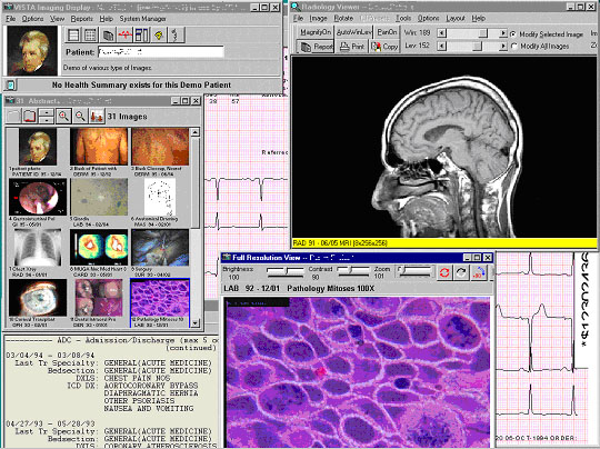
The major goal of VISTA Imaging is to facilitate medical decision-making by delivering complete multimedia patient information to the clinician's desktop in an integrated manner. Windows-based workstations, which are interfaced to the main hospital system in a client-server architecture, make images and associated text data available at all times anywhere in the hospital.
VISTA Imaging handles high quality image data from many specialties, including cardiology, pulmonary and gastrointestinal medicine, pathology, radiology, hematology, and nuclear medicine. VISTA Imaging can also process textual reports from the hospital information system, scanned documents and electrocardiograms.
VISTA Imaging is integrated with the Computerized Patient Record System (CPRS) to provide a comprehensive electronic patient record. VISTA Imaging improves the quality of patient care, enhances clinicians' communication, and is used routinely for daily work, conferences, morning reports, educational seminars, and during ward rounds.
VISTA Imaging's diagnostic display software (VISTARad) can be used for the filmless interpretation of radiology studies and for radiology workflow management.
VISTA Imaging is made up of the following components:
- Core Infrastructure
- Document Imaging
- Filmless Radiology
- Imaging Ancillary Systems
Each component is described in the following pages.
Features
VISTA Imaging:
- Provides display, manipulation, and management functions for a wide variety of medical images such as radiographs, sonograms, EKG tracings, gastroenterology studies, pulmonary bronchoscope exams, podiatry, dermatology and ophthalmology images. Images can be any sort of multimedia data, including digital images, motion video clips, graphics, scanned documents, and audio files.
- Is integrated with CPRS, allowing users to view images automatically for a selected patient. When a user views a radiology report or progress note in CPRS, the associated images are easily available.
- Provides an interface between VISTA and commercial PACS systems using DICOM Gateway software.
- Automatically acquires complete studies from DICOM-compliant modalities (CT, MRI, digital x-ray, ultrasound, etc), associates the studies with the correct patient and report, and stores the studies in VISTA Imaging for inclusion in the electronic patient record.
- Manages image file storage, management, and retrieval from magnetic and optical disk servers and supports data capture, storage, and retrieval over a local or wide area network (WAN).
- Provides access to electronic medical records from remote VA medical facilities over the VA intranet, if appropriate access privileges are assigned.
VistA Imaging: Core Infrastructure
Status: VistA Imaging: missing - Alternate Available
![]() Not VistA Imaging: missing - Uses proprietary components not available outside the VA
Not VistA Imaging: missing - Uses proprietary components not available outside the VA
![]() Alt O3 DPACS IHE compliant from the O3 Consortium - Open Source - Java based
Alt O3 DPACS IHE compliant from the O3 Consortium - Open Source - Java based
Overview
The Core Imaging Infrastructure includes the components used to capture, store, and display all types of images. Images can be captured using video cameras, digital cameras, document and color scanners, x-ray scanners, and files created by commercial capture software. Images can also be directly acquired from DICOM-compliant devices such as CT scanners, MR scanners and digital x-ray machines.
Captured images are accessible through VISTA Imaging or the Computerized Patient Record System (CPRS) graphical user interface (GUI) on the clinician's desktop. Viewing a procedure/exam report or progress note in CPRS allows the user to automatically access any associated images.
Core Imaging Infrastructure is made up of the following:
- DICOM text gateways, which provide patient and order information to medical devices (such as CT scanners and digital radiography systems), allowing selection of the examination to be performed. The data provided by DICOM text gateways complies with the DICOM Modality Worklist standard. DICOM text gateways can also be used to communicate patient and order information to commercial PACS (Picture Archiving and Communications Systems).
- DICOM image gateways, which allow VISTA Imaging to receive images from PACS systems or acquisition devices. Image gateways can also be used to transfer images from the VISTA system to any DICOM-compliant storage devices for display, printing, or teleradiology purposes.
- Windows-based workstation software for image display and capture.
- The Background Processor, which manages image storage on various network devices, including magnetic storage (RAID) and optical storage (jukebox) as a long-term archive.
- The VISTA Imaging database, which manages the links between images and patient electronic records.
- The commercially available equipment required by VISTA Imaging, including magnetic servers, optical disk jukeboxes, and utility workstations.
Features
The Core Imaging Infrastructure:
- Acquires images and multimedia data.
- Stores images to allow immediate access and long-term permanent storage.
- Communicates and displays images in a timely manner.
- Processes different types of images from different specialties.
- Links images to VistA's integrated patient record so that they can be retrieved by patient or study/progress note.
- Protects security and privacy of images, and prevents alteration of images after capture.
- Enables remote viewing and capture of images.
- Uses non-proprietary hardware.
VistA Imaging: Document Imaging
Status: VistA Imaging: missing - Alternate Available
![]() Not VistA Imaging: missing - Uses proprietary components not available outside the VA
Not VistA Imaging: missing - Uses proprietary components not available outside the VA
![]() Alt O3 DPACS IHE compliant from the O3 Consortium - Open Source - Java based
Alt O3 DPACS IHE compliant from the O3 Consortium - Open Source - Java based
Overview
Document Imaging allows scanned and electronically generated documents to be associated with the online patient record and displayed on clinical workstations. The benefits of Document Imaging include:
- Online availability of all information in the electronic patient record, including handwritten papers, drawings, signed documents, and medical correspondence.
- Linkage of paper-based patient information to the electronic patient record, making all patient information quickly available and easily retrievable from a single source.
- Immediate availability of critical documents, such as advance directives and informed consent forms, at the time of need.
- Elimination of lost or misfiled medical chart information.
- Potential savings due to reduced medical records staff and file room costs. Possible benefits include decreased retrieval, filing and delivery functions, reduced volume of paper records, and faster retrieval time for clinicians.
Document Imaging will also provide interfaces to commercial document scanning systems and systems that generate documents electronically.
Features
- Scanning and indexing of black-and-white, grayscale, and color documents, including: signed advance directives, consent forms, annotated drawings, outside medical records documents, and administrative documents such as Means Test forms.
- Automatic transmission of signed means test documents to the Health Eligibility Center (HEC), in compliance with VA requirements.
- Document image storage in short and long term storage devices.
- Display of images for clinical and administrative purposes.
- Printing of document images.
VistA Imaging: Filmless Radiology
Status: VistA Imaging: missing - Alternate Available
![]() Not VistA Imaging: missing - Uses proprietary components not available outside the VA
Not VistA Imaging: missing - Uses proprietary components not available outside the VA
![]() Alt O3 DPACS IHE compliant from the O3 Consortium - Open Source - Java based
Alt O3 DPACS IHE compliant from the O3 Consortium - Open Source - Java based
Overview
Filmless Radiology uses high-resolution workstations and high-speed servers to allow radiology departments to operate without generating x-ray film. Workstations running VISTARad, VISTA Imaging's diagnostic image display software, are used by radiologists for the online interpretation of images acquired by CR, CT, MRI, and other modalities. VISTARad is integrated with the VistA Hospital Information System and with the Radiology Package in particular.
Features
- Exam lists, which are based on exam status or patient name, are used to divide the pool of available exams into useful categories. Exam lists are created using information from the Radiology package and the VISTA patient database.
- Site-specific custom lists can be created to reflect the workflow of individual departments.
- In addition to images, other parts of a patient's medical record, such as laboratory and pathology information, can be displayed using the VISTA Health Summary reporting capability.
- A full range of image display and manipulation features for radiology and nuclear medicine images are provided.
- Multiple exams can be displayed concurrently, allowing for comparisons with prior studies.
- Exam locks are used to prevent duplication of effort.
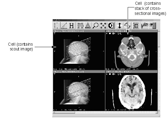
- Specialized display tools are provided for CT, MRI, and angiography exams, including cine viewing, series linking, and series synchronization.
VistA Imaging: Imaging Ancillary Systems
Status: VistA Imaging: missing - Alternate Available
![]() Not VistA Imaging: missing - Uses proprietary components not available outside the VA
Not VistA Imaging: missing - Uses proprietary components not available outside the VA
![]() Alt O3 DPACS IHE compliant from the O3 Consortium - Open Source - Java based
Alt O3 DPACS IHE compliant from the O3 Consortium - Open Source - Java based
Overview
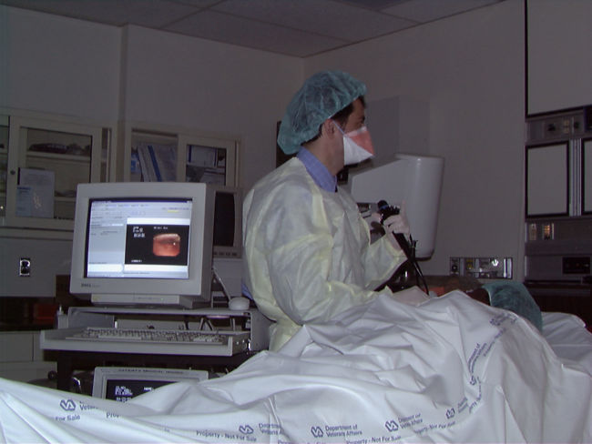
VistA Imaging's Ancillary Systems component captures, stores, and displays images for a particular service or specialty. This may be accomplished using the Clinical Capture workstation or by interfaces to commercial systems. These commercial systems are typically small systems, and often incorporate special-purpose hardware. They often have components that assist clinicians in creating exam or procedure reports.
Features
- Allows direct capture of images from a variety of sources, such as standard video outputs from medical devices, and digital cameras.
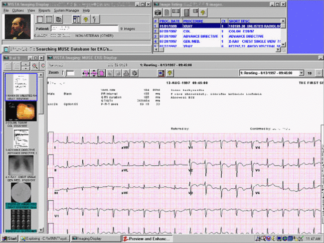
- Interfaces to a commercial EKG system for display of electrocardiograms on clinical workstations.
- Supports automatic DICOM interfaces for capture of specialty images from compliant systems (DICOM Modality Worklist Conformance Requirements are provided to sites purchasing specialty equipment).
- In the future DICOM and Clinical Workstation support for ophthalmology, dental, endoscopy, pathology, cardiology, and other specialties will be included.
- Support for acquisition of DICOM Structured Report information is planned for the future.


