Microscope: Difference between revisions
(Added details derived from the video snapshot.) |
Maraschino (talk | contribs) |
||
| (8 intermediate revisions by 5 users not shown) | |||
| Line 1: | Line 1: | ||
{{stub}} |
|||
== Overview - The $1 video microscope == |
== Overview - The $1 video microscope == |
||
| Line 16: | Line 14: | ||
Some pixels are visibly rectangular, suggesting perhaps ~1/3 pixel resolution, or about 50 um. |
Some pixels are visibly rectangular, suggesting perhaps ~1/3 pixel resolution, or about 50 um. |
||
A 50 um resolution would mean you could see amoebas (about the size of a pixel), and large plant cells, but most animal cells would be fuzzy dots (about 1/10 of a pixel). A human hair is slightly narrower than a pixel. |
A 50 um resolution would mean you could see amoebas (about the size of a pixel), and large plant cells, but most animal cells would be fuzzy dots (about 1/10 of a pixel). A human hair is slightly narrower than a pixel. |
||
[http://www.vendian.org/howbig/?&page=micropaper These two] |
|||
[http://www.vendian.org/howbig/?&page=microfloor pages] might provide context. |
|||
The XO screen pixels are slightly smaller than the big red circle ("your screen dot") halfway down the first page. |
|||
== Techniques == |
== Techniques == |
||
| Line 28: | Line 30: | ||
As [http://microscopy.fsu.edu/optics/intelplay/qx3darkfield.html here]. |
As [http://microscopy.fsu.edu/optics/intelplay/qx3darkfield.html here]. |
||
== Other |
== Other Microscopes == |
||
| ⚫ | |||
=== Veho 004 === |
|||
Full specifications on this site: http://www.veho-uk.com/main/shop_detail.aspx?article=40&mode=specifications |
|||
Just as a minor clarification on the specifications, the microscope does not have a range magnification of 20X-to-400X. |
|||
In actuality (and from multiple user reviews) this microscope has "2" magnifications 20X and 400X. The current range numbers on the barrel of the device is misleading. For a detailed customer review click [http://www.amazon.com/review/R1IGLAHE0T8Y8L/ref=cm_cr_rdpvoterdr#R1IGLAHE0T8Y8L.2115.Helpful.Reviews here]. |
|||
The following instruction is summary of an e-mail from Kevin Gordon. This microscope is still under development to run using "guvcview" on XO. For this current installation, we will use "Cheese" as it is known to work reliably with it. |
|||
What is Cheese? Cheese is a Linux program that allows your microscope (or other webcams and USB camera/video devices) to work with your XO to take photos and videos and apply special effects on them. In order to make your microscope work with Cheese it has to work Gstreamer Framework and Video4Linux2 (V4L2) or Video4Linux (V4L). For more information on Cheese visit their site [http://projects.gnome.org/cheese/ here]. |
|||
==== with an XO 1.5 ==== |
|||
1. Plug in your Veho 004 microscope to one of the USB ports on your XO. Power up your XO. |
|||
2. On the "Home" view. Right-click on the "Terminal" activity. Make sure you have a good internet connection. |
|||
3. You have to be a root user to start the installation. Type this at the prompt: |
|||
<br /> |
|||
su |
|||
4. To install Cheese. Type the following at the prompt: |
|||
yum install cheese |
|||
5. You will get an indication that the installation is done. You are now ready to run cheese. To do so, exit the root mode by typing at the the # prompt: |
|||
exit |
|||
at the % prompt type: |
|||
cheese |
|||
and voila! You are now ready for a fun learning experience with your microscope! |
|||
=== Intel Play USB Microscope === |
|||
| ⚫ | |||
== News == |
|||
*[http://laptop.media.mit.edu/laptopnews.nsf/2e76a5a80bc36cbf85256cd700545fa5/3e24474967102e7285257357005e048f?OpenDocument OLPC News 2007-09-15] 3. Microscope: Professor Robert Shapiro visited Mary Lou Jepsen at OLPC last week to discuss more issues of optimal microscope design to allow the XO to provide diagnosis of HIV/AIDs, TB, and malaria, which kill more than six-million people every year, worldwide. Low-cost detection of these diseases could save many lives. Surprisingly, the key for detection is not high magnification; low magnification of a large image area and a dye coupled with violet-colored LEDs for illumination can be combined with image processing is sufficient. Professor Shapiro showed a prototype microscope to Mary Lou and discussed the basic requirements. Barrett Comiskey (whose has been designing a periscope) is also working on a low-cost microscope for the XO. |
|||
[[Category:Hardware ideas]] |
|||
[[Category:Peripherals]] |
|||
[[Category:Science]] |
|||
Latest revision as of 18:47, 6 March 2011
Overview - The $1 video microscope
With additional lenses, the XO can be used as a video microscope. A prototype $1 lens exists.
A video of Mary Lou's prototype microscope attachment for the XO video camera. She compares the display of an XO and a more typical ThinkPad.
The microscope, which is said to have ~ 100× magnification, could be useful to analyzing water quality, among other things.
Details
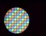
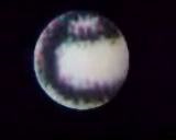
From the youtube video, it looks like the field of view is about 9.5 pixels, so 1.2 mm (because the screen is 200 ppi). Pixels are about 130 um diagonally. Some pixels are visibly rectangular, suggesting perhaps ~1/3 pixel resolution, or about 50 um. A 50 um resolution would mean you could see amoebas (about the size of a pixel), and large plant cells, but most animal cells would be fuzzy dots (about 1/10 of a pixel). A human hair is slightly narrower than a pixel.
These two pages might provide context. The XO screen pixels are slightly smaller than the big red circle ("your screen dot") halfway down the first page.
Techniques
Magnification is often only the first step in being able to see details.
Aside from staining, a variety of techniques might be adapted to the XO. QX3 techniques suggests some possibilities.
Dark field microscopy
- Also known as "darkfield microscopy".
In direct sunlight, with a small square of black construction paper, it might be possible to do a form of dark field microscopy. As here.
Other Microscopes
Veho 004
Full specifications on this site: http://www.veho-uk.com/main/shop_detail.aspx?article=40&mode=specifications
Just as a minor clarification on the specifications, the microscope does not have a range magnification of 20X-to-400X. In actuality (and from multiple user reviews) this microscope has "2" magnifications 20X and 400X. The current range numbers on the barrel of the device is misleading. For a detailed customer review click here.
The following instruction is summary of an e-mail from Kevin Gordon. This microscope is still under development to run using "guvcview" on XO. For this current installation, we will use "Cheese" as it is known to work reliably with it.
What is Cheese? Cheese is a Linux program that allows your microscope (or other webcams and USB camera/video devices) to work with your XO to take photos and videos and apply special effects on them. In order to make your microscope work with Cheese it has to work Gstreamer Framework and Video4Linux2 (V4L2) or Video4Linux (V4L). For more information on Cheese visit their site here.
with an XO 1.5
1. Plug in your Veho 004 microscope to one of the USB ports on your XO. Power up your XO.
2. On the "Home" view. Right-click on the "Terminal" activity. Make sure you have a good internet connection.
3. You have to be a root user to start the installation. Type this at the prompt:
su
4. To install Cheese. Type the following at the prompt:
yum install cheese
5. You will get an indication that the installation is done. You are now ready to run cheese. To do so, exit the root mode by typing at the the # prompt:
exit
at the % prompt type:
cheese
and voila! You are now ready for a fun learning experience with your microscope!
Intel Play USB Microscope
The XO has also been used with the now-discontinued $100 Intel Play QX3 USB Microscope.[1]
News
- OLPC News 2007-09-15 3. Microscope: Professor Robert Shapiro visited Mary Lou Jepsen at OLPC last week to discuss more issues of optimal microscope design to allow the XO to provide diagnosis of HIV/AIDs, TB, and malaria, which kill more than six-million people every year, worldwide. Low-cost detection of these diseases could save many lives. Surprisingly, the key for detection is not high magnification; low magnification of a large image area and a dye coupled with violet-colored LEDs for illumination can be combined with image processing is sufficient. Professor Shapiro showed a prototype microscope to Mary Lou and discussed the basic requirements. Barrett Comiskey (whose has been designing a periscope) is also working on a low-cost microscope for the XO.

