Microscope
Overview - The $1 video microscope
With additional lenses, the XO can be used as a video microscope. A prototype $1 lens exists.
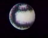
A video of Mary Lou's prototype microscope attachment for the XO video camera. She compares the display of an XO and a more typical ThinkPad.
The microscope, which is said to have ~ 100× magnification, could be useful to analyzing water quality, among other things.
Details
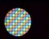
From the youtube video, it looks like the field of view is about 9.5 pixels, so 1.2 mm (because the screen is 200 ppi). Pixels are about 130 um diagonally. Some pixels are visibly rectangular, suggesting perhaps ~1/3 pixel resolution, or about 50 um. A 50 um resolution would mean you could see amoebas (about the size of a pixel), and large plant cells, but most animal cells would be fuzzy dots (about 1/10 of a pixel). A human hair is slightly narrower than a pixel.
These two pages might provide context. The XO screen pixels are slightly smaller than the big red circle ("your screen dot") halfway down the first page.
Techniques
Magnification is often only the first step in being able to see details.
Aside from staining, a variety of techniques might be adapted to the XO. QX3 techniques suggests some possibilities.
Dark field microscopy
- Also known as "darkfield microscopy".
In direct sunlight, with a small square of black construction paper, it might be possible to do a form of dark field microscopy. As here.
Other microscopes
The XO has also been used with the now-discontinued $100 Intel Play QX3 USB Microscope.[1]
News
- OLPC News 2007-09-15 3. Microscope: Professor Robert Shapiro visited Mary Lou Jepsen at OLPC last week to discuss more issues of optimal microscope design to allow the XO to provide diagnosis of HIV/AIDs, TB, and malaria, which kill more than six-million people every year, worldwide. Low-cost detection of these diseases could save many lives. Surprisingly, the key for detection is not high magnification; low magnification of a large image area and a dye coupled with violet-colored LEDs for illumination can be combined with image processing is sufficient. Professor Shapiro showed a prototype microscope to Mary Lou and discussed the basic requirements. Barrett Comiskey (whose has been designing a periscope) is also working on a low-cost microscope for the XO.

